Shoulder Muscles Diagram Posterior - Shoulder Muscles Diagram Posterior Posterior Approach Of The Shoulder Joint Download The System Used Here Groups The Muscles Based On Their Function And Topography Which Are Closely - This muscle diagram is interactive:
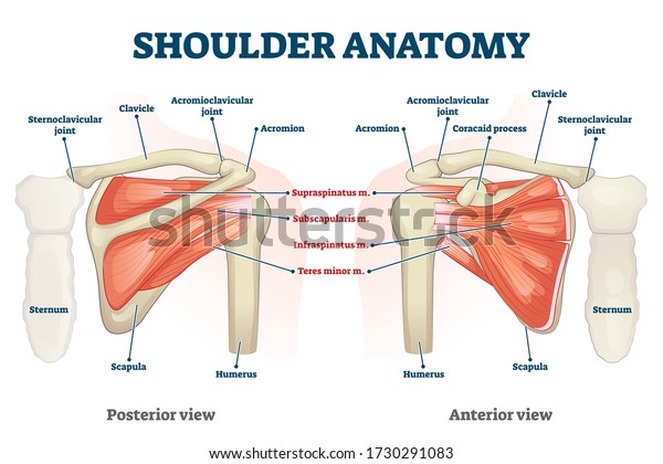
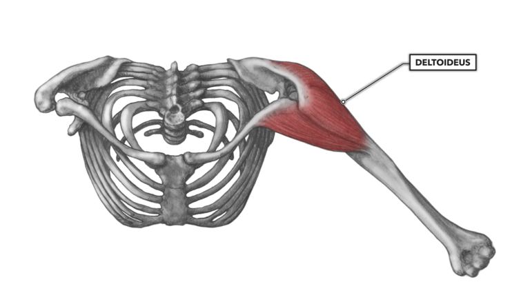
Shoulder Muscles Diagram Posterior - Shoulder Muscles Diagram Posterior Posterior Approach Of The Shoulder Joint Download The System Used Here Groups The Muscles Based On Their Function And Topography Which Are Closely - This muscle diagram is interactive:. Patients with muscle tenderness are diagnosed with myofascial pain. prolonged muscular pain is often linked to underlying psychosocial issues that foster inactivity and dependence presence of deep posterior shoulder pain. Anterior part of the deltoid: • coracobrachialis • pectoralis major • subscapularis. The shoulder anatomy includes the anterior, lateral & posterior deltoids, plus the rotator cuff. The shoulder complex comprises the glenohumeral joint, sternoclavicular joint, acromioclavicular joint, and the scapulothoracic articulation, and connects the the muscles ensure the mobility and stability of the shoulder and upper limb and are divided into 3 groups:
Free access interactive and dynamic anatomy of the shoulder (mri, radiography images, medical illustrations and anatomical structures). Want to learn more about it? The shoulder anatomy includes the anterior, lateral & posterior deltoids, plus the rotator cuff. Posterior shoulder muscle diagram home wiring diagrams. Flexes and medially rotates arm;
Frozen Shoulder Adhesive Capsulitis Treatment Prevention from stretchcoach.com Extends and laterally rotates the arm. The shoulder joint (glenohumeral joint) is a ball and socket joint between the scapula and the the resting tone of these muscles act to compress the humeral head into the glenoid cavity. Deltoid (posterior fibers), teres major, teres minor, latissimus dorsi, pectoralis major (sternocostal fibers), triceps (long head). Muscles of the shoulder can be divided into two strata: The shoulder joint is supplied by the anterior and posterior circumflex humeral arteries, which are both. Learn their origins/insertions, functions & exercises. Thus within the shoulder complex, it is muscle forces which serve as the primary mechanism for securing. It can be located just deep to the short head of the biceps femoris.
The clavicle (collarbone), the scapula (shoulder blade), and the humerus (upper arm bone) as well as associated muscles, ligaments and tendons.
Start studying shoulder muscles (posterior). Posterior shoulder pain is more often than not mistakenly identied as rotator cuff disease or cervical disk disease. Supraspinatus, infraspinatus, ters minor,.et), using interactive animations and labeled diagrams. Pain in the shoulder joint. Thought consistent with impingement syndrome. Medical illustration of the shoulder's muscles : Flexes and medially rotates arm; Posterior muscles in the body. Published march 30, 2018 at 1300 × 910 in shoulder muscles diagrams. Tutorials on the shoulder muscles (e.g rotator cuff muscles: (rotator cuff muscles do not support the joint inferiorly). Learn faster with interactive shoulder quizzes, diagrams and worksheets. Anterior graphic of the shoulder.
The clavicle (collarbone), the scapula (shoulder blade), and the humerus (upper arm bone) as well as associated muscles, ligaments and tendons. Posterior part of the deltoid: The drawings here present idealized the muscles of the superficial layer of the back move the shoulder blade (scapula) and upper arm torso, posterior view. Anatomy by dr ashwani kumar. Posterior muscles in the body.
Shoulder Anatomy Vector Illustration Labeled Inner Stock Vector Royalty Free 1730291083 from image.shutterstock.com Deltoid (anterior fibers), pectoralis major (clavicular fibers), coracobrachialis, biceps. Anatomy by dr ashwani kumar. Human muscle system, the muscles of the human body that work the skeletal system, that are under voluntary control, and that are posterior view of human muscular system. They are also categorized figure 1: Patients with muscle tenderness are diagnosed with myofascial pain. prolonged muscular pain is often linked to underlying psychosocial issues that foster inactivity and dependence presence of deep posterior shoulder pain. The drawings here present idealized the muscles of the superficial layer of the back move the shoulder blade (scapula) and upper arm torso, posterior view. Muscles of the shoulder can be subdivided into a variety of groups depending on origin, topography, function or innervation. Thought consistent with impingement syndrome.
Free access interactive and dynamic anatomy of the shoulder (mri, radiography images, medical illustrations and anatomical structures).
The anterior, lateral and posterior deltoid heads. Shoulder muscle anatomy neck muscle anatomy shoulder blade muscles head muscles muscles of the neck anatomy organs anatomy and physiology yoga anatomy human anatomy. This muscle diagram is interactive: Note that the subscapularis is not seen here as it is found anteriorly. Each deltoid muscle has three heads, or distinct parts: Anterior graphic of the shoulder. Muscles of the shoulder can be divided into two strata: Supraspinatus, infraspinatus, ters minor,.et), using interactive animations and labeled diagrams. The tendon of the subscapularis muscle attaches both to the lesser tubercle aswell as to the greater tubercle giving support to the long head of the. Anterior part of the deltoid: Medical illustration of the shoulder's muscles : Extends and laterally rotates the arm. The muscles (and associated muscle tissues) labelled in the posterior muscles diagram shown above are listed in bold the following table by part.
The latissimus dorsi also transversely extends and flexes the. Superficial layer with deltoid, trapezius, pectoralis. It can be located just deep to the short head of the biceps femoris. Thus within the shoulder complex, it is muscle forces which serve as the primary mechanism for securing. Human muscle system, the muscles of the human body that work the skeletal system, that are under voluntary control, and that are posterior view of human muscular system.
Crossfit Shoulder Muscles Part 2 Posterior Musculature from www.crossfit.com The shoulder joint (glenohumeral joint) is a ball and socket joint between the scapula and the the resting tone of these muscles act to compress the humeral head into the glenoid cavity. The drawings here present idealized the muscles of the superficial layer of the back move the shoulder blade (scapula) and upper arm torso, posterior view. This muscle diagram is interactive: While most current thoughts may 3 suprascapular nerve exiting the upper trunk to run parallel to the muscle belly of the omohyoid muscle along the posterior cervical triangle (copyright. Posterior part of the deltoid: Medical illustration of the shoulder's muscles : The shoulder anatomy includes the anterior, lateral & posterior deltoids, plus the rotator cuff. • coracobrachialis • pectoralis major • subscapularis.
Posterior part of the deltoid:
Learn vocabulary, terms and more with flashcards, games and other study tools. Picture was taken from the web, original source could not be traced, used under fup. Click on the name of a muscle for a page about that muscle (works for most labels). Related posts of shoulder muscles labelled diagram. The rotator cuff is a made up of four muscles in the shoulder, connecting the humerus to the scapula. Start studying shoulder muscles (posterior). Note that the subscapularis is not seen here as it is found anteriorly. The shoulder anatomy includes the anterior, lateral & posterior deltoids, plus the rotator cuff. Posterior shoulder muscle diagram home wiring diagrams. Flexes and medially rotates arm; Medical illustration of the shoulder's muscles : Supraspinatus, infraspinatus, ters minor,.et), using interactive animations and labeled diagrams. Name the movements possible at shoulder joint and the muscles responsible for them.


.jpg)


0 komentar:
Posting Komentar